Endovenous laser
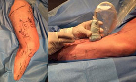
After performing an ultrasound to map the patient's specific anatomy and varicose veins, the leg is prepped and draped for a sterile technique. Under light sedation, the procedure begins with local anesthesia to numb the skin. Guided by ultrasound, the saphenous vein is cannulated.
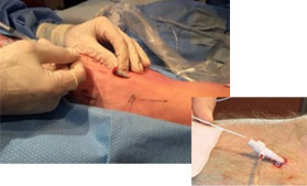
Next, a laser fiber is advanced along the entire length of the incompetent saphenous vein, guided by ultrasound.
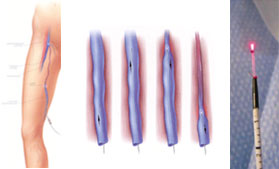
Once everything is ready, the optical fiber is activated and slowly withdrawn (1 mm per second). A heat point is emitted from the tip of the laser fiber, sealing the saphenous vein, and the blood flow is automatically redirected to the surrounding normal veins.
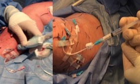
To conclude, the surface varicose veins, which were already cannulated using small venous access catheters at the beginning of the treatment (a technique developed by Dr. Danylewick), are now ready for sclerosing injections.
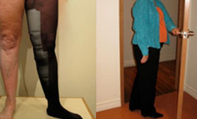
A compression stocking is put on to aid healing, and the patient leaves the clinic pain-free, able to resume activities quickly.
